

SUM Ultimate Medicare OPD: 9:30 AM – 5:30 PM (Mon–Sat)
DR
Dr. Radhamadhab SahuSenior Consultant & HOD · ENT & Skull Base
Patient Education & Clinical Literacy

Patient Resources & Guides

Empowering patients and families with verified medical knowledge. These educational articles are written from an evidence-based clinical perspective to help you understand your symptoms and prepare for specialist consultations.

Rhinology & Sinus Health5 min read • Educational Guide

Understanding Chronic Sinusitis: Causes, Symptoms & Modern Care

What causes paranasal sinus blockage, when medical therapies suffice, and how Functional Endoscopic Sinus Surgery (FESS) restores natural sinus ventilation without external incisions.

The human skull contains four paired paranasal sinus air cavities: maxillary, ethmoid, frontal, and sphenoid. In healthy individuals, these produce mucus that traps dust and pathogens, which micro-cilia constantly sweep through narrow ostia into the nasal cavity.

When chronic inflammation, allergy, or anatomical obstruction blocks these narrow drainage pathways, mucus stagnates. Bacteria or fungi proliferate, leading to persistent facial heaviness, purulent nasal discharge, hyposmia (loss of smell), and chronic headaches.

While initial episodes are treated medically with saline irrigations and intranasal steroid sprays, refractory cases lasting beyond 12 weeks often benefit from Functional Endoscopic Sinus Surgery (FESS). FESS uses optical cameras through the nostrils to widen natural drainage channels while preserving healthy mucosa.

Clinical Pearls & Key Takeaways

  • Symptoms lasting longer than 12 weeks indicate chronic rhinosinusitis, requiring formal endoscopic evaluation.
  • Routine over-the-counter decongestant nasal drops (e.g. xylometazoline) should NEVER be used for more than 5 consecutive days to prevent rhinitis medicamentosa (rebound congestion).
  • Modern FESS utilizes dissolvable bio-absorbable spacers, avoiding painful traditional packing removal.
Understanding Chronic Sinusitis: Causes, Symptoms & Modern Care
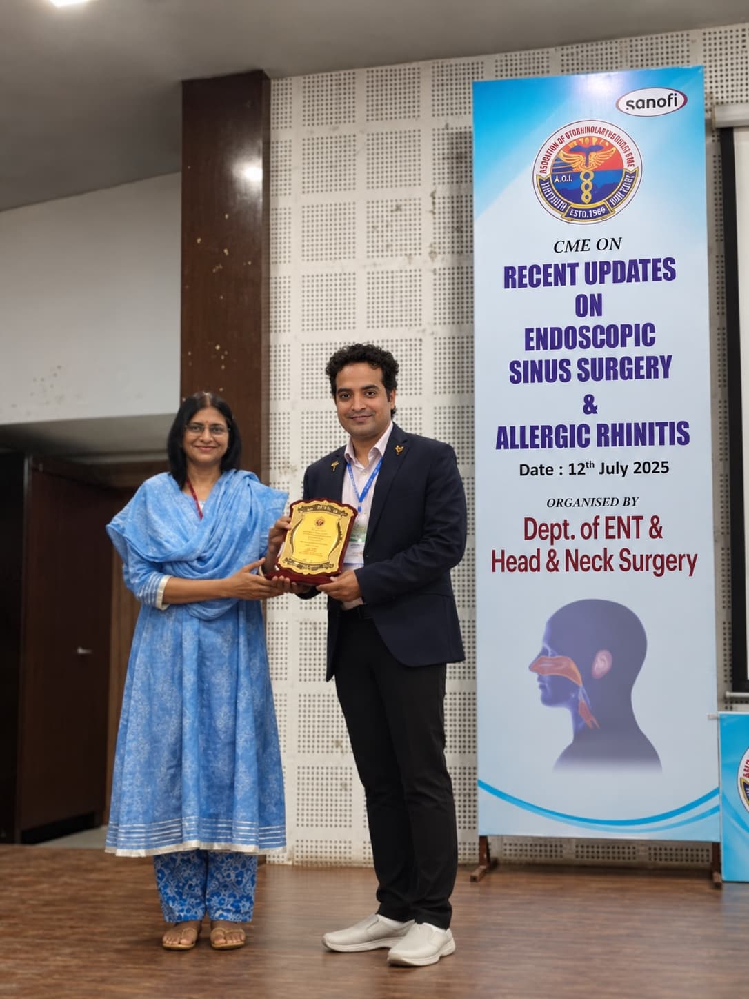
Sinus Surgery CME

Dr. Radhamadhab Sahu honored on stage at the AOI State CME on Endoscopic Sinus Surgery (FESS) & Allergic Rhinitis.

Have Symptoms of this Condition?

Evaluations are conducted with advanced high-definition endoscopy at SUM Ultimate Medicare and Dr. Radhamadhab ENT and Cancer Clinic.

Schedule Clinical Review →

Medical Reviewer: Dr. Radhamadhab Sahu (Senior Consultant & HOD)Schedule an Evaluation for this condition →
Nasal Airway4 min read • Educational Guide

Deviated Nasal Septum (DNS) & Chronic Nasal Obstruction

How a deviated midline septum disrupts nasal airflow, causes unilateral congestion, and how endoscopic septoplasty straightens the cartilage with minimal recovery time.

The nasal septum is the cartilage and bone partition separating the left and right nasal airways. In many individuals, trauma during birth, childhood sports, or natural developmental spurts causes the septum to deviate significantly to one side.

A deviated septum restricts laminar airflow, forcing mouth-breathing, dry mouth on waking, disrupted sleep, snoring, and compensatory enlargement (hypertrophy) of the opposite inferior turbinate.

Endoscopic Septoplasty is a minimally invasive surgical procedure performed entirely inside the nostrils. Under microscopic magnification, the surgeon straightens and re-aligns the deviated cartilage without any external marks, bruising, or alterations to the external shape of the nose.

Clinical Pearls & Key Takeaways

  • Septoplasty is an internal functional procedure; it does not change the external shape of your nose unless combined with rhinoplasty.
  • Most patients resume light desk work within 3 to 5 days following endoscopic septoplasty.
Deviated Nasal Septum (DNS) & Chronic Nasal Obstruction
Airway Surgery Faculty

Dr. Radhamadhab Sahu delivering an invited lecture on endoscopic septoplasty and airway reconstruction corridors.

Have Symptoms of this Condition?

Evaluations are conducted with advanced high-definition endoscopy at SUM Ultimate Medicare and Dr. Radhamadhab ENT and Cancer Clinic.

Schedule Clinical Review →
Medical Reviewer: Dr. Radhamadhab Sahu (Senior Consultant & HOD)Schedule an Evaluation for this condition →
Clinical Guidance4 min read • Educational Guide

When Should You Consult an ENT Specialist? 7 Red Flag Symptoms

Learn which ear, nose, throat, and neck symptoms warrant prompt specialist evaluation rather than continued self-medication.

Many mild ENT symptoms resolve within a week or two. However, certain persistent or progressive signs indicate underlying structural, neurological, or neoplastic processes requiring expert otolaryngology investigation.

1. Persistent Hoarseness: Any change in voice lasting more than 3 weeks requires video laryngoscopy to examine the vocal folds.

2. Unilateral Clear Watery Nasal Discharge: Fluid that drips from only one nostril when bending forward may represent cerebrospinal fluid (CSF leak) from the skull base.

3. Asymmetric Hearing Loss or Sudden Deafness: Sudden loss of hearing in one ear is a medical emergency requiring immediate steroids within 48 to 72 hours for best recovery.

4. Unilateral Ear Pain with Normal Ear Canal: Referred otalgia may originate from an underlying tonsillar, tongue base, or laryngeal pathology.

5. Painless Firm Neck Lump: Any new lump in the neck that persists beyond 2 to 3 weeks must be evaluated with ultrasound and fine needle aspiration cytology (FNAC).

6. Severe Snoring with Breathing Pauses: Choking awakenings and unrefreshing sleep signify Obstructive Sleep Apnea, which stresses cardiovascular function.

7. Chronic Ear Drainage: Foul-smelling ear discharge suggests chronic suppurative otitis media or cholesteatoma requiring surgical eradication.

Clinical Pearls & Key Takeaways

  • Early consultation prevents chronic complications and allows less invasive endoscopic interventions.
When Should You Consult an ENT Specialist? 7 Red Flag Symptoms
Consultation Chamber

Distinctive head-silhouette awards wall and surgical honours inside Dr. Radhamadhab Sahu's clinic in Bhubaneswar.

Have Symptoms of this Condition?

Evaluations are conducted with advanced high-definition endoscopy at SUM Ultimate Medicare and Dr. Radhamadhab ENT and Cancer Clinic.

Schedule Clinical Review →
Medical Reviewer: Dr. Radhamadhab Sahu (Senior Consultant & HOD)Schedule an Evaluation for this condition →
Otology & Hearing6 min read • Educational Guide

Understanding Ear Symptoms: Perforations, Cholesteatoma & Implants

A guide to eardrum repair (tympanoplasty), cholesteatoma risks, and how cochlear and bone-anchored hearing aids restore auditory connection.

The ear is divided into the external canal, the middle ear cleft containing the three smallest bones in the human body (malleus, incus, and stapes), and the delicate fluid-filled inner ear (cochlea).

Chronic ear infections often cause a hole (perforation) in the tympanic membrane (eardrum). A perforation compromises hearing and permits water and bacteria to enter the middle ear, triggering recurrent drainage. Tympanoplasty reconstructs the eardrum using natural graft tissue (temporalis fascia or tragal cartilage), sealing the ear and restoring hearing.

When bone-eroding skin cyst tissue (cholesteatoma) develops inside the mastoid air cells, it can erode the hearing bones and facial nerve. Timely mastoidectomy eradicates this disease and preserves inner ear function.

For individuals with profound sensorineural deafness non-responsive to hearing aids, modern Cochlear Implants bypass damaged hair cells to stimulate the auditory nerve directly.

Clinical Pearls & Key Takeaways

  • Never let water enter an ear with a known eardrum perforation; use cotton soaked in petroleum jelly during showers.
  • Simultaneous same-day bilateral tympanoplasty can repair both ears in a single sitting under expert hands.
Understanding Ear Symptoms: Perforations, Cholesteatoma & Implants
Verified Clinical Follow-Up

Long-term hearing restoration: Patient Abhishek Rao with Dr. Sahu at 2-year post-mastoidectomy follow-up.

Have Symptoms of this Condition?

Evaluations are conducted with advanced high-definition endoscopy at SUM Ultimate Medicare and Dr. Radhamadhab ENT and Cancer Clinic.

Schedule Clinical Review →
Medical Reviewer: Dr. Radhamadhab Sahu (Senior Consultant & HOD)Schedule an Evaluation for this condition →
Skull Base Surgery6 min read • Educational Guide

What is Endoscopic Skull Base Surgery? A Patient's Primer

How surgeons access deep pituitary tumors, clival chordomas, and cranial base leaks through the nostrils without opening the skull.

The skull base is the complex floor of the cranial cavity supporting the brain and pierced by critical cranial nerves and major arteries. Historically, reaching tumors located here required large craniotomies, external facial incisions, and extensive brain retraction.

Over the past two decades, high-definition optical endoscopes have revolutionized skull base surgery. By entering through the nostrils and traversing the sphenoid sinus, surgeons gain direct panoramic access to the sella turcica (pituitary gland), clivus, and anterior cranial fossa.

Using optical neuronavigation, micro-doppler probes, and specialized angled instruments, tumors like pituitary macroadenomas and chordomas are removed with high precision, minimizing blood loss and hospital stay.

Clinical Pearls & Key Takeaways

  • Endoscopic endonasal approaches leave zero external facial incisions.
  • Post-operative care emphasizes strict avoidance of nose-blowing or heavy lifting to allow the skull base flap to heal securely.
What is Endoscopic Skull Base Surgery? A Patient's Primer
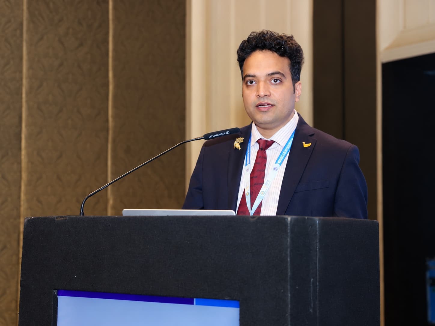
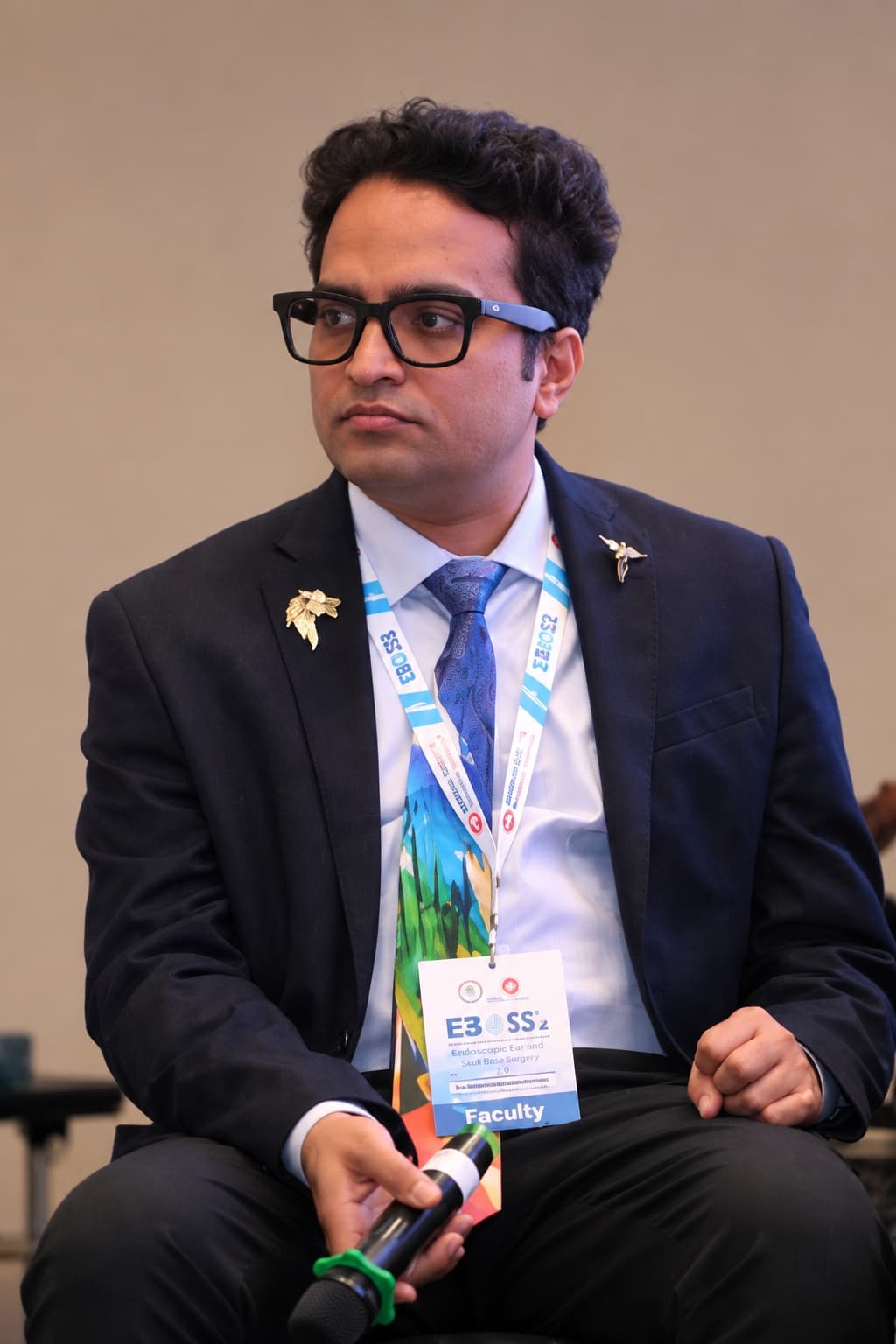
EBOSS 2.0 Faculty

Dr. Radhamadhab Sahu serving as expert faculty at EBOSS 2.0 (Endoscopic Ear & Skull Base Surgery).

Have Symptoms of this Condition?

Evaluations are conducted with advanced high-definition endoscopy at SUM Ultimate Medicare and Dr. Radhamadhab ENT and Cancer Clinic.

Schedule Clinical Review →
Medical Reviewer: Dr. Radhamadhab Sahu (Senior Consultant & HOD)Schedule an Evaluation for this condition →
Consultation & Appointments

Schedule Your Clinical Consultation

Dr. Radhamadhab Sahu consults across two authorized clinical centers in Kalinga Nagar, Bhubaneswar. Choose your preferred care setting below to connect directly with the respective appointment desk.

Quaternary Hospital OPD

SUM Ultimate Medicare

Department of ENT and Skull Base Surgery

Satellite GPS Map of SUM Ultimate Medicare, Ghatikia
K-8, Ghatikia Main RoadGPS Map

Primary venue for comprehensive outpatient evaluations, in-hospital endoscopies, neurosurgical skull base surgeries, and inpatient surgical care.

K-8, Kalinga Nagar, Ghatikia, Bhubaneswar, Odisha 751003
Consultation Hours: 9:30 AM – 5:30 PM (Monday – Saturday; Closed Sundays & Public Holidays)
Private Clinic

E.N.T. & Cancer Super-Speciality Clinic

Skull Base Surgery Head & Neck Cancer Center

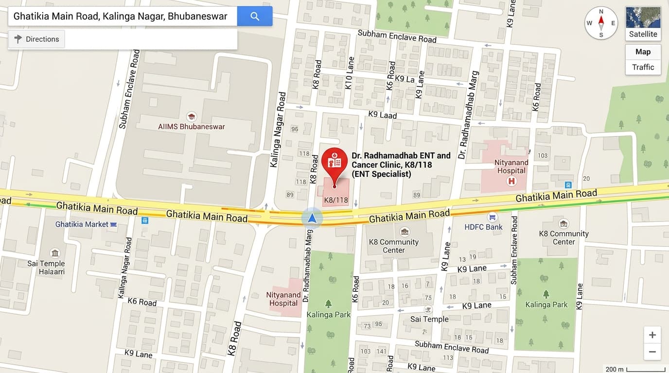
Realistic Road Map of E.N.T. & Cancer Super-Speciality Clinic, Ghatikia
Plot K8/118, Ghatikia Main RoadStreet Map

Dedicated private clinic for personalized ENT assessments, endoscopic examinations, post-operative surgical reviews, and specialized skull base consultations.

K8/118, Ghatikia Main Road, Kalinga Nagar, Bhubaneswar, Odisha 751003
Consultation Hours:Mon – Sat: 8:00 AM – 11:00 AM & 6:00 PM – 9:00 PMSunday: 8:00 AM – 12:30 PM
Important Guidelines for Your Visit:

To ensure thorough evaluation, please bring all previous medical records, recent blood reports, radiological imaging films/CDs (CT scans or MRI scans of the brain, sinuses, or neck), and any existing audiometry / hearing tests.

Book Visit